File:Myoviruses P-SSM2 and P-SSM4.gif
Myoviruses_P-SSM2_and_P-SSM4.gif (600 × 412 pixels, file size: 200 KB, MIME type: image/gif)
Summary
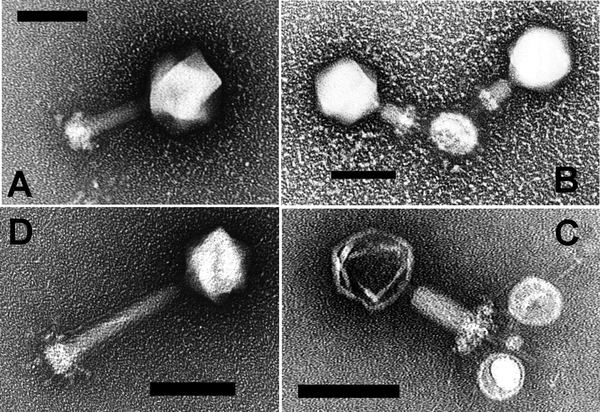
Electron Micrograph of Negative-Stained Prochlorococcus Myoviruses P-SSM2 and P-SSM4
Myovirus P-SSM2 with (A) non-contracted tail and (B) contracted tail, and myovirus P-SSM4 with (C) contracted tail and (D) non-contracted tail. Note the T4-like capsid, baseplate, and tail structure in both myoviruses. Scale bars indicate 100 nm.
From: Three Prochlorococcus Cyanophage Genomes: Signature Features and Ecological Interpretations Sullivan MB, Coleman ML, Weigele P, Rohwer F, Chisholm SW PLoS Biology Vol. 3, No. 5, e144 doi:10.1371/journal.pbio.0030144.
Licensing
This media, Myoviruses P-SSM2 and P-SSM4.gif, is licenced under the Creative Commons Attribution 2.5 Unported License
You are free:
To Share — To copy, distribute and transmit the work; To Remix — To adapt the work.
Under the following conditions:
Attribution — You must attribute the work in the manner specified by the author or licensor (but not in any way that suggests that they endorse you or your use of the work).
For any reuse or distribution, you must make clear to others the licence terms of this work (the best way to do this is with a link to this licence's web page). Any of the above conditions can be waived if you get permission from the copyright holder. Nothing in this licence impairs or restricts the author's moral rights.
Read the full licence.
File history
Click on a date/time to view the file as it appeared at that time.
| Date/Time | Thumbnail | Dimensions | User | Comment | |
|---|---|---|---|---|---|
| current | 19:53, 11 March 2022 | | 600 × 412 (200 KB) | Maintenance script (talk | contribs) | == Summary == Importing file |
You cannot overwrite this file.
File usage
The following 3 pages use this file: