File:Hagmann 2008 Mapping the Structural Core of Human Cerebral Cortex Fig. 1.png: Difference between revisions
Jump to navigation
Jump to search
imported>Daniel Mietchen ({{Image_Details |description = Extraction of a Whole Brain Structural Connectivity Network. (1) High-resolution T1 weighted and diffusion spectrum MRI (DSI) is acquired. DSI is represented with a zoom on the axial slice of the reconstructed diffusion map, showing an orientation distribution function at each position represented by a deformed sphere whose radius codes for diffusion intensity. Blue codes for the head-feet, red for left-right, and green for anterior-posterior orientations. (2)...) |
(== Summary == Importing file) Tag: Server-side upload |
||
| Line 1: | Line 1: | ||
== Summary == | == Summary == | ||
Importing file | |||
Latest revision as of 19:53, 11 March 2022
Summary
Importing file
File history
Click on a date/time to view the file as it appeared at that time.
| Date/Time | Thumbnail | Dimensions | User | Comment | |
|---|---|---|---|---|---|
| current | 19:53, 11 March 2022 | 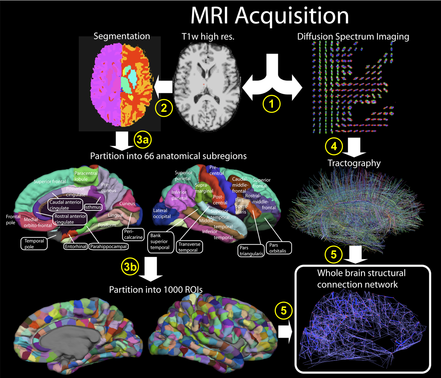 | 1,689 × 1,446 (1.7 MB) | Maintenance script (talk | contribs) | == Summary == Importing file |
You cannot overwrite this file.
File usage
The following page uses this file: