File:Fmri visual.jpg: Difference between revisions
Jump to navigation
Jump to search
imported>Richard Pettitt ({{Image notes |Description=3-D fMRI map of regions in the brain that exhibit blood flow changes in response to visual stimulation. Scientists are using powerful MRI machines which allow, noninvasively, for the localization and visualization of active brain regions. Functional MRI (fMRI), a technique that measures small and localized blood flow changes that follow increase in neuronal activity in the brain, was done on a ultra-high field magnet (9.4 Tesla) using high spatial resolution (0.15 x...) |
imported>Caesar Schinas m (Replace Pd-USgov by PD-USgov) |
||
| Line 10: | Line 10: | ||
|Other versions=}} | |Other versions=}} | ||
== Licensing/Copyright status == | == Licensing/Copyright status == | ||
{{ | {{PD-USgov}} | ||
Revision as of 03:00, 28 May 2009
File history
Click on a date/time to view the file as it appeared at that time.
| Date/Time | Thumbnail | Dimensions | User | Comment | |
|---|---|---|---|---|---|
| current | 19:54, 11 March 2022 | 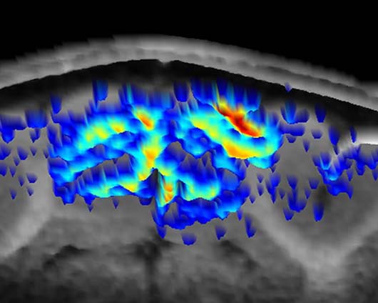 | 530 × 424 (43 KB) | Maintenance script (talk | contribs) | == Summary == Importing file |
You cannot overwrite this file.
File usage
The following page uses this file: