Cortical variability: Difference between revisions
Jump to navigation
Jump to search

imported>Daniel Mietchen (image legend) |
imported>Daniel Mietchen (Image:Brain-variability-STS.png does not display properly any more) |
||
| (4 intermediate revisions by the same user not shown) | |||
| Line 1: | Line 1: | ||
{{subpages}} | {{subpages}} | ||
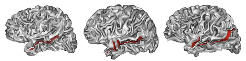
{{Image|Brain-variability-STS.png|right| | {{Image|Brain-variability-STS-horizontal.png|right|500px|[[Gyrification|Cortical folding pattern]] of the [[human brain]], highlighting the [[anatomical variability]] of the [[superior temporal sulcus]], which may consist of one (left), two (middle) or three (right) parts. Note that other cortical structures also exhibit considerable variation.}} | ||
'''Cortical variability''' describes the [[anatomical variability]] of the [[cerebral cortex]]. It manifests itself in variations of the size, shape or number of cortical structures, e.g. the number or shape of [[sulcus (neuroanatomy)|sulci]] and [[gyri]], and is typically expressed in terms of the [[statistical variation]] of [[grey matter volume]], [[cortical thickness]], [[gyrification]], [[cortical surface area]], [[cortical connectivity]] or related [[brain morphometry|brain morphometric]] measures across a set of [[brain]]s or [[brain images]]. | |||
The term is usually used in reference to normal healthy adults but it can also refer to other groups of subjects, including patients with [[brain disorder]]s. Besides the health state, cortical variability is a function of multiple factors, including [[age]], [[gender]], [[ethnicity]] and [[species (biology)|species]]. | |||
Latest revision as of 19:36, 2 May 2010

(CC) Image: Lefèvre and Mangin, 2010
Cortical folding pattern of the human brain, highlighting the anatomical variability of the superior temporal sulcus, which may consist of one (left), two (middle) or three (right) parts. Note that other cortical structures also exhibit considerable variation.
Cortical folding pattern of the human brain, highlighting the anatomical variability of the superior temporal sulcus, which may consist of one (left), two (middle) or three (right) parts. Note that other cortical structures also exhibit considerable variation.
Cortical variability describes the anatomical variability of the cerebral cortex. It manifests itself in variations of the size, shape or number of cortical structures, e.g. the number or shape of sulci and gyri, and is typically expressed in terms of the statistical variation of grey matter volume, cortical thickness, gyrification, cortical surface area, cortical connectivity or related brain morphometric measures across a set of brains or brain images.
The term is usually used in reference to normal healthy adults but it can also refer to other groups of subjects, including patients with brain disorders. Besides the health state, cortical variability is a function of multiple factors, including age, gender, ethnicity and species.