Chordoma: Difference between revisions
Jump to navigation
Jump to search


imported>Daniel Mietchen (+image) |
imported>Daniel Mietchen (image legend) |
||
| Line 7: | Line 7: | ||
At least one [[susceptibility gene]] has been identified<ref name=Yang2009>{{CZ:Ref:Yang 2009 T (brachyury) gene duplication confers major susceptibility to familial chordoma}}</ref> but currently, no therapy is in sight, leaving affected patients with an average life expectancy of five to ten years after diagnosis. | At least one [[susceptibility gene]] has been identified<ref name=Yang2009>{{CZ:Ref:Yang 2009 T (brachyury) gene duplication confers major susceptibility to familial chordoma}}</ref> but currently, no therapy is in sight, leaving affected patients with an average life expectancy of five to ten years after diagnosis. | ||
{{Image|Chordoma immunohistochemistry.png|right|350px|Different immunohistochemical markers of chondroma. | {{Image|Chordoma immunohistochemistry.png|right|350px|Different immunohistochemical markers of chondroma. A: E-E, × 5; chordoma composed of nests and cords of tumour cells; B: E-E, × 20; [[physaliphorous cell]]s containing multiple clear [[cytoplasm]]ic [[vacuole]]s; C: S-100, × 20; [[immunohistochemistry]] showing staining for [[S-100 protein]]; D: CK, × 20; immunohistochemistry showing staining for [[cytokeratin]]; E: EMA, × 20; immunohistochemistry showing staining for [[Epithelial membrane antigen|EMA]]; F: Vimentin, × 20; immunohistochemistry showing staining for [[Vimentin]].}} | ||
==References== | ==References== | ||
<references/> | <references/> | ||
Revision as of 11:03, 12 May 2010

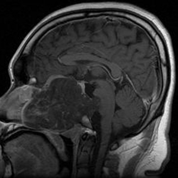
A chordoma (also known as choroid meningioma, chordocarcinoma, chordoepithelioma, or notochordoma) is a malignant tumor arising from the embryonic remains of the notochord.[1]
Such malformations can occur anywhere in the bony tissue surrounding the spinal cord, though typically its mobile parts at the cranial or caudal end. The prevalence is about 1 in a million in the United States.
At least one susceptibility gene has been identified[2] but currently, no therapy is in sight, leaving affected patients with an average life expectancy of five to ten years after diagnosis.

(CC) Photo: Larizza et al., 2005
Different immunohistochemical markers of chondroma. A: E-E, × 5; chordoma composed of nests and cords of tumour cells; B: E-E, × 20; physaliphorous cells containing multiple clear cytoplasmic vacuoles; C: S-100, × 20; immunohistochemistry showing staining for S-100 protein; D: CK, × 20; immunohistochemistry showing staining for cytokeratin; E: EMA, × 20; immunohistochemistry showing staining for EMA; F: Vimentin, × 20; immunohistochemistry showing staining for Vimentin.
Different immunohistochemical markers of chondroma. A: E-E, × 5; chordoma composed of nests and cords of tumour cells; B: E-E, × 20; physaliphorous cells containing multiple clear cytoplasmic vacuoles; C: S-100, × 20; immunohistochemistry showing staining for S-100 protein; D: CK, × 20; immunohistochemistry showing staining for cytokeratin; E: EMA, × 20; immunohistochemistry showing staining for EMA; F: Vimentin, × 20; immunohistochemistry showing staining for Vimentin.
References
- ↑ Anonymous (2024), Chordoma (English). Medical Subject Headings. U.S. National Library of Medicine.
- ↑ Yang XR, Ng D, Alcorta DA, Liebsch NJ, Sheridan E, Li S et al. (2009). "T (brachyury) gene duplication confers major susceptibility to familial chordoma". Nat Genet 41 (11): 1176-8. DOI:10.1038/ng.454. PMID 19801981. Research Blogging. [e]
- Based on comparative genomic hybridization data from four affected families, variations in the 6q27 region of the human chromosome 6 were found to be correlated with chordoma, thus suggesting the T gene as a potential susceptibility gene for the disease.