Photobacterium phosphoreum: Difference between revisions
imported>Fatima Giron |
imported>Fatima Giron |
||
| Line 87: | Line 87: | ||
P. phosphoreum is particularly preferred for laboratory culture and isolation. It can be extracted from dead seafish, squid or octopus. It grows at low temperature (10-15˚C). It is cultivated in complex media like “BOSS.” The BOSS medium can be made of NaCl, glycerol, peptone like Bacto-Peptone and beef extract diluted in water. Another medium that you can use is luminescence medium (LM) which contains glycerol, yeast extract, tryptone, CaC''O''<sub>3</sub> and agar [http://www.disknet.com/indiana_biolab/b203.htm]. | P. phosphoreum is particularly preferred for laboratory culture and isolation. It can be extracted from dead seafish, squid or octopus. It grows at low temperature (10-15˚C). It is cultivated in complex media like “BOSS.” The BOSS medium can be made of NaCl, glycerol, peptone like Bacto-Peptone and beef extract diluted in water. Another medium that you can use is luminescence medium (LM) which contains glycerol, yeast extract, tryptone, CaC''O''<sub>3</sub> and agar [http://www.disknet.com/indiana_biolab/b203.htm]. | ||
They have a high growth rate. You can visualize the glowing colonies formed on the plate in a dark room. P. phosphoreum is a good to practice isolation because it is easy to culture and is not pathogenic [http://www.biology.pl/bakterie_sw/bac_dyd_en.html]. | They have a high growth rate. You can visualize the glowing colonies formed on the plate in a dark room. P. phosphoreum is a good bioluminescent bacterium to practice isolation because it is easy to culture and is not pathogenic [http://www.biology.pl/bakterie_sw/bac_dyd_en.html]. | ||
We can be observed the morphology of these bioluminescent bacteria under the [[light microscope]] with the aid of some [[staining methods]]. The more preferable are the [[gram stain]] and the [[negative staining]] [http://www.biology.pl/bakterie_sw/bac_mf_en.html]. | We can be observed the morphology of these bioluminescent bacteria under the [[light microscope]] with the aid of some [[staining methods]]. The more preferable are the [[gram stain]] and the [[negative staining]] [http://www.biology.pl/bakterie_sw/bac_mf_en.html]. | ||
Revision as of 18:18, 7 May 2009
For the course duration, the article is closed to outside editing. Of course you can always leave comments on the discussion page. The anticipated date of course completion is May 21, 2009. One month after that date at the latest, this notice shall be removed. Besides, many other Citizendium articles welcome your collaboration! |
| Photobacterium phosphoreum | ||||||||||||||
|---|---|---|---|---|---|---|---|---|---|---|---|---|---|---|
 | ||||||||||||||
| Scientific classification | ||||||||||||||
| ||||||||||||||
| Binomial name | ||||||||||||||
| Photobacterium phosphoreum |
Description and significance
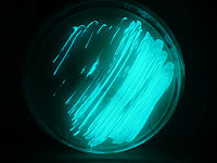
Photobacterium phosphoreum is a luminescent bacterium. It is a straight rod and gram-negative bacterium with a large size and round shape. It is motile. It can grow at a low temperature around 4˚C, but no at 35˚C or above. It emits the brightest light from all bioluminescent bacteria.[1] Its optimum temperature is at 18-25˚C .[2] This bacterium produces a blue-green light due to the catalytic activity of luciferase. The light, that P. phosphoreum produces, is known as cold light because the light producing reaction doesn't require and generate much heat.[3] It is oxidase positive because they can use D-glucose as their principle carbon source.[4] It can be cultivated in Long and Hammer Agar (1% NaCl). It is known as a symbiotic bacterium that lives in the light organ of some marine fishes. It can also live freely in seawater, saprophytically and parasitically.[5]
Its major significance is their symbiotic relationship with some marine animals like fishes and squids. These marine animals have light organs that provide P. phosphoreum bacteria a safe place to inhabit and obtain food; while they use the light that the bacteria provide for camouflage, communication and even for attracting mates or escape from predators.[6] Light emission can also aid to the propagation of the host. Another role that P. phosphoreum has is its ability of signaling the relative toxicity of a substance. This can happen due to the connection of the light producing process with its cellular metabolism. If the toxins disrupt the cellular metabolism, then the strength of the light produced decreases.[2]
Genome structure
Photobacterium phosphoreum major lineage is Gammaproteobacteria. It contains a single circular chromosome. The genome size is about 4.0 Mb. It has a G+C content of genomic DNA of 41%. The number of genes are 4576. The coding percent is 84.0%. It can be isolated by using plating methods Microbial Genome Sequencing Project.
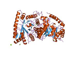
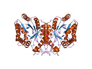
P. phosphoreum contains in its genome a lux gene that codes for the enzyme luciferase. This enzyme transforms chemical energy into light energy. Luciferase is a heterodimer with alpha and beta subunits. These two subunits are coded by luxA and luxB respectively.
Cell structure and metabolism
P. phosphoreum belongs to the phylum Proteobacteria. All bacteria that belong to this phylum are known to be Gram-negative. As gram-negative P. phosphoreum contain an outer membrane made of lipopolysaccharide outside and inside phospholipids, a periplasma space, a thin peptidoglycan layer and finally the plasma membrane Encyclopedia of Life Sciences. They are straight rod with 0.8-1.3 um in diameter and 1.8-2.4 um in length. P. phosphoreum has 1-3 unsheathed polar flagella [1].
P. phosphoreum is a chemoorganotroph which is capable of respiratory and fermentative metabolism [2]. It is a facultative anaerobe that can grow in the absence of oxygen when appropriate electron-acceptors are present. It doesn’t denitrify; in other word, it cannot use nitrogen molecules [3].
P. phosphoreum can produce blue-green light with the help of an enzyme called luciferase. “Luciferase catalyzes the reaction and uses reduced flavin mononucleotide, molecular oxygen, and a long-chain aldehyde as substrate” (Willey, Sherwood and Woolverton 559). These substrates are known as bacterial luciferin.
Equation
FMNH2 + O2 + RCHO + luciferase → FMH + H2 + RCOOH + light
When the excited, enzyme-bound flavin intermediate moves to ground state, it emits the light which is one of the products of the reaction [4]. "The bioluminescence quantum yield has been estimated to be 10–30%" Encyclopedia of Life Sciences. If you have observed in the photosynthetic equation reaction, oxygen is not produce therefore P. phosphoreum is known to have an anoxygenic photosynthesis Encyclopedia of Life Sciences.
Ecology
P. phosphoreum is mostly considered a marine bacterium because sodium ions are required for its growth. It lives in the depth of the ocean, seawater, marine sediments, in the guts of marine animals, and on the surface of decomposing fish. When they are dispersed they produce a very weak light; however when they are very close to each other they produce light efficiently. P. phosphoreum acts as the light bulb in the in a dark environment like in the depth of the ocean Biology.
Cultivation and Isolation
P. phosphoreum is particularly preferred for laboratory culture and isolation. It can be extracted from dead seafish, squid or octopus. It grows at low temperature (10-15˚C). It is cultivated in complex media like “BOSS.” The BOSS medium can be made of NaCl, glycerol, peptone like Bacto-Peptone and beef extract diluted in water. Another medium that you can use is luminescence medium (LM) which contains glycerol, yeast extract, tryptone, CaCO3 and agar [5].
They have a high growth rate. You can visualize the glowing colonies formed on the plate in a dark room. P. phosphoreum is a good bioluminescent bacterium to practice isolation because it is easy to culture and is not pathogenic [6].
We can be observed the morphology of these bioluminescent bacteria under the light microscope with the aid of some staining methods. The more preferable are the gram stain and the negative staining [7].
Application to Biotechnology
P. phosphoreum due to its bioluminescence ability is use in some biotechnology processes. The enzyme luciferase, found in P. phosphoreum, can be use together with NAD(P)H dehydrogenase to estimate the concentration of NAD(P)H Oxford Dictionary of Biochemistry and Molecular Biology. This enzyme can be also use for determining the pollution of water and toxicities. The lux gene, which codes for luciferase, can be integrated in the genome of another bacteria in order to trace visually the bacterial population development. “Because luminescence can occur over and over again and because a bacterium's cycle of luminescence is very short (i.e., a cell is essentially blinking on and off), luminescence allows a near instantaneous (i.e., "real time") monitoring of bacterial behavior” (Wilmoth and Lee p354) [8].
Current Research
Biogenic amine formation and bacterial contribution in fish, squid and shellfish
This study was done by Min-Ki Kim, Jae-Hyung Mah and Han-Joon Hwang. Their main concern was whether engineered nanoparticles (NPs) can become a possible risk to human health and the environment. To test the toxicity produce by nanoparticles, in this case a stable metal (Au), bactericide (Ag) and Fe3O4, Photobacterium phosphoreum, germination test and other anaerobic toxicity tests were used. P. phosphoreum can be isolated from fish, squid and shellfish. In all tests, the results showed low or zero toxicity. Furthers studies are still being perform Department of Food and Biotechnology.
Sentinels of the seas
This research is from a journal named New Scientist and the author is Andy Coghlan. It was discovered that Photobacterium phosphoreum HE-1a can sense water pollution. The glowing colony got dimmer when pollutants were present. Researchers have discovered that colonies of glowing bacteria get dimmer when they sense pollution. It responded to almost every pollutant; however, it couldn’t distinguish them. The researcher concluded that “HE-1a could be used as an early warning system” that would indicate the presence of pollution in water. We can keep an eye on waste container discarded in the ocean (Coghlan p. 21) [9].
Riboflavin synthesis genes are linked with the lux operon of Photobacterium phosphoreum
Lee, O'Kane and Meighen have shown that riboflavin synthesis genes and lux genes are linked to each other. Four gene after the lux operon and luxG were identify. They code for 3,4-dihydroxy-2-butanone 4-phosphate synthetase, GTP cyclohydrolase II, riboflavin synthetase, and lumazine synthetase (Lee, O'Kane and Meighen p. 2100-4) [10].
Photocatalytic disinfection of marine bacteria using fluorescent light
This study was done by Leung TY, Chan CY, Hu C, Yu JC and Wong PK from Chinese University of Hong Kong. “Photocatalytic oxidation (PCO) using fluorescent light was used to disinfect two marine bacteria: Alteromonas alvinellae and Photobacterium phosphoreum” [11]. P. phosphoreum was less susceptibility to PCO than A. alvinellae due to their fatty acid molecules and levels of superoxide dismutase (SOD) and catalase (CAT). The PCO treatment is a irreversible making the strains of the tested bacteria inactivate [12].
References
- ↑ Eddleman, Harold, Ph. D. Isolation of Pure Cultures Of Bacteria April 2009
- ↑ 2.0 2.1 Bunch, Joshua. Photobacterium phosphoreum: A Microbial Flashlight. April 2009
- ↑ Wilson, Tracy V. How Bioluminescence Works. How Stuff Work April 2009
- ↑ Willey, Joanne, Linda Sherwood and Christopher Woolverton. Prescott, Harley, and Klein's Microbiology. 7th ed. New York: New York, 2008. pg 557
- ↑ Budsberg, K.J.; C.F. Wimpee & J.F. Braddock (2003), "Isolation and identification of Photobacterium phosphoreum from an unexpected niche: migrating …", Applied and Environmental Microbiology 69 (11): 6938–6942. Retrieved on 2009-04-29
- ↑ http://mrw.interscience.wiley.com/emrw/9780470015902/els/article/a0003064/current/html Only opens in QC
Bioluminescent Bacteria. April 2009
Brenda Wilmoth Lerner and K. Lee. World of Microbiology and Immunology. Lerner. Vol. 1. Detroit: Gale, 2003.
Coghlan, Andy. (June 1998) Sentinels of the seas. New Scientist v. 158 no. 2137 p. 21 Wilson Web Journal
Hagström, Åke. Photobacterium sp. SKA34. Microbial Genome Sequencing Project April 2009.
Hastings, Woodland J and Kurt L Krause (January 2007) doi: 10.1038/npg.els.0003064
Kersters, Karel et al. (April 2006) doi: 10.1038/npg.els.0004312
Kima, Min-Ki, Jae-Hyung Mahb and Han-Joon Hwang. (2009) doi:10.1016/j.foodchem.2009.02.010
Lee, Chan Yong, Dennis J. O'Kane and Edward A. Meighen. (April 1994) Riboflavin synthesis genes are linked with the lux operon of Photobacterium phosphoreum. J Bacteriol 176(7):2100-4 PMID: 8144477
Leung et al. (Sept 2008) Photocatalytic disinfection of marine bacteria using fluorescent light. Water Res. 42(19):4827-37 PMID: 18842281
Madanecki, Piotr. Luminescent Bacteria Website April 2009
Nealson, Kenneth (January 2002). Luminescent Bacteria: An Introduction. April 2009
Photobacterium phosphoreum April 2009
Skerman VBD et al. (1980) Photobacterium phosphoreum April 2009

