File:DTI-sagittal-fibers.jpg: Difference between revisions
imported>Caesar Schinas m (Robot: Changing template: GFDL) |
imported>Caesar Schinas m (Bot: Replace Template:Image_notes_* with Template:Image_Details) |
||
| Line 1: | Line 1: | ||
== Summary == | == Summary == | ||
{{ | {{Image_Details | ||
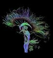
| | | description = English: Visualization of a DTI measurement of a human brain. Depicted are reconstructed fiber tracts that run through the mid-sagittal plane. Especially prominent are the U-shaped fibers that connect the two hemispheres through the corpus callosum (the fibers come out of the image plane and consequently bend towards the top) and the fiber tracts that descend toward the spine (blue, within the image plane). | ||
Deutsch: Traktographie-Verfahren rekonstruieren aus den Messdaten der Diffusions-Tensor-Bildgebung den anzunehmenden Verlauf größerer Nervenbahnen. Hier dargestellt sind die Ergebnisse für ein menschliches Gehirn; um die Übersichtlichkeit zu wahren, beschränkt sich die Abbildung auf Bahnen, die die Medianebene schneiden. Insbesondere sind dies die U-förmigen Faserbündel, die die beiden Hirnhälften verbinden (sie durchstoßen die Bildebene und sind nach oben gebogen) sowie die Faserbündel, die zum Rückenmark ziehen (blau dargestellt, liegen innerhalb der Bildebene). | Deutsch: Traktographie-Verfahren rekonstruieren aus den Messdaten der Diffusions-Tensor-Bildgebung den anzunehmenden Verlauf größerer Nervenbahnen. Hier dargestellt sind die Ergebnisse für ein menschliches Gehirn; um die Übersichtlichkeit zu wahren, beschränkt sich die Abbildung auf Bahnen, die die Medianebene schneiden. Insbesondere sind dies die U-förmigen Faserbündel, die die beiden Hirnhälften verbinden (sie durchstoßen die Bildebene und sind nach oben gebogen) sowie die Faserbündel, die zum Rückenmark ziehen (blau dargestellt, liegen innerhalb der Bildebene). | ||
| | | author = Thomas Schultz {{#if:http://www.sci.utah.edu/~gk/DTI-data/|(http://www.sci.utah.edu/~gk/DTI-data/)}} | ||
| copyright = Thomas Schultz | |||
| | | source = http://en.wikipedia.org/wiki/Image:DTI-sagittal-fibers.jpg | ||
| | | date-created = September 22, 2006 | ||
| | | pub-country = | ||
| | | notes = Rendering is own work, using a modified version of the BioTensor application developed at the University of Utah. The dataset is courtesy of Gordon Kindlmann at the Scientific Computing and Imaging Institute, University of Utah, and Andrew Alexander, W.M. Keck Laboratory for Functional Brain Imaging and Behaviour, University of Wisconsin, Madison. | ||
| versions = | |||
}} | |||
== Licensing/Copyright status == | == Licensing/Copyright status == | ||
{{GNU|FDL|1.2|+}} | {{GNU|FDL|1.2|+}} | ||
Revision as of 01:54, 22 June 2009
Summary
| Title / Description
|
English: Visualization of a DTI measurement of a human brain. Depicted are reconstructed fiber tracts that run through the mid-sagittal plane. Especially prominent are the U-shaped fibers that connect the two hemispheres through the corpus callosum (the fibers come out of the image plane and consequently bend towards the top) and the fiber tracts that descend toward the spine (blue, within the image plane).
Deutsch: Traktographie-Verfahren rekonstruieren aus den Messdaten der Diffusions-Tensor-Bildgebung den anzunehmenden Verlauf größerer Nervenbahnen. Hier dargestellt sind die Ergebnisse für ein menschliches Gehirn; um die Übersichtlichkeit zu wahren, beschränkt sich die Abbildung auf Bahnen, die die Medianebene schneiden. Insbesondere sind dies die U-förmigen Faserbündel, die die beiden Hirnhälften verbinden (sie durchstoßen die Bildebene und sind nach oben gebogen) sowie die Faserbündel, die zum Rückenmark ziehen (blau dargestellt, liegen innerhalb der Bildebene). |
|---|---|
| Author(s)
|
Thomas Schultz (http://www.sci.utah.edu/~gk/DTI-data/) |
| Copyright holder
|
Thomas Schultz See below for license/re-use information. |
| Source
|
http://en.wikipedia.org/wiki/Image:DTI-sagittal-fibers.jpg |
| Date created
|
September 22, 2006 |
| Country of first publication
|
Please edit this page and add the country of first publication, or note that it is unknown. |
| Notes
|
Rendering is own work, using a modified version of the BioTensor application developed at the University of Utah. The dataset is courtesy of Gordon Kindlmann at the Scientific Computing and Imaging Institute, University of Utah, and Andrew Alexander, W.M. Keck Laboratory for Functional Brain Imaging and Behaviour, University of Wisconsin, Madison. |
| Other versions
|
If there are other versions of this media on CZ, please list them here. |
| Using this image on CZ
|
Copy the code below to add this image to a Citizendium article, changing the size, alignment, and caption as necessary.
|
Please send email to manager A T citizendium.org .
Licensing/Copyright status
DTI-sagittal-fibers.jpg is licenced under the GNU Free Documentation License v1.2 or later
Permission is granted to copy, distribute and/or modify this document under the terms of the GNU Free Documentation License, version 1.2 or any later version published by the Free Software Foundation; with no Invariant Sections, no Front-Cover Texts, and no Back-Cover Texts.
Read the licence for full details
Notice of CC-by-sa incompatibility
Text on the Citizendium is licensed under the Creative Commons Attribution ShareAlike licence.
This media may not be used under those or closely similar terms without separate permission of the copyright holder, or the user's own fair use determination.
File history
Click on a date/time to view the file as it appeared at that time.
| Date/Time | Thumbnail | Dimensions | User | Comment | |
|---|---|---|---|---|---|
| current | 18:57, 11 March 2022 |  | 1,021 × 1,125 (203 KB) | Maintenance script (talk | contribs) | == Summary == Importing file |
You cannot overwrite this file.
File usage
The following 3 pages use this file: