Vibrio cholerae: Difference between revisions
imported>Zerla Lau |
imported>Zerla Lau |
||
| Line 65: | Line 65: | ||
==References== | ==References== | ||
http://en.wikipedia.org/wiki/Vibrio_cholerae [http://en.wikipedia.org/wiki/Vibrio_cholerae] | http://en.wikipedia.org/wiki/Vibrio_cholerae [http://en.wikipedia.org/wiki/Vibrio_cholerae] | ||
Revision as of 21:59, 16 April 2008
Articles that lack this notice, including many Eduzendium ones, welcome your collaboration! |
 | ||||||||||||||
| Scientific classification | ||||||||||||||
| ||||||||||||||
| Binomial name | ||||||||||||||
| Vibrio cholerae |
Description and significance
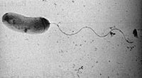
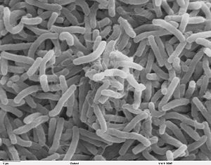
Vibrio Cholerae is a gram-negative, curved rod-shaped bacterium that has a single polar flagellum. It is an aerobic organism and thrives best in alkaline mediums.[1]
Genome structure
The Cholera genome sequencing started in 1997 and was completed several years ago which revealed the bacterium`s two circular chromosomes with the larger of the chromosome having approximately 3 million base pairs. The smaller of the pair chromosomes have approximately one million. Of the total 3,885 genes of Vibrio cholerae, approximately 75% of those genes reside on the larger chromosome. Obtaining this extra chromosome gives a competitive advantage for the survival of V. cholerae in diverse environments. [2]
According to the authors Kaper et. al at the University of Maryland School of Medicine, Vibrio cholerae`s familial species seized a large plasmid and over time the two organisms exchanged genes and genetic information and relied on each other for survival.
Cell structure and metabolism
Vibrio cholerae generate three polysaccharides that relate to the cell surface. They are lipopolysaccharide, capsule, and rugose exopolysaccharide. The lipopolysaccharide and the capsule both assist in the avoidance of host defense mechanisms. The rugose polysaccharide helps sustain the bacterium in aquatic environments when nutrients are poor and insufficient.[3]
Ecology
Vibrio cholerae resides in aquatic habitats and are able to thrive in both fresh water and marine environments. In fresh water environments, the bacteria will inhabit lakes, ponds, and rivers. Marine environments such as oceans and seas are also places where V. cholerae can be found.
V. cholerae are also readily found in costal areas where there are significant amounts of algal bloom. Overabundance of algal blooms such as phytoplankton and zooplankton have tremendous consequences on food and water safety because Vibrio cholerae attaches itself to small marine crustaceans called Copepods which help spread Cholera.[4]
Pathology
Vibrio cholerae was first isolated by an Italian scientist, Filippo Pacini, after the Asiatic Cholera Pandemic of 1846- 63 swept through Italy. Pacini accurately described the disease an acute loss of fluid and electrolytes caused by the bacteria acting on the epithelial intestinal mucuosa. However, despite having published numerous publications on his research and dicoveries, his scientific work was rejected and ignored by the community due to the prevailing Miasma Theory of Disease which scientists thought was the valid explanation for the cause of Cholera. Thirty years later in 1884,a German physician Robert Koch, who was one of the founders of Microbiology became the acknowledged discoverer of V. cholerae. Koch was oblivious to the research done by Pacini and since Koch was well regarded in the scientic community, he became of the acknowleged discoverer of V. cholerae.[5]
Vibrio cholerae is the causative agent of a serious epidemic disease Cholera. Cholera is a harmful bacterial ailment that often causes severe diarrhea which is usually procurred by the ingestion of food or water contaminated with V. cholerae. In epidemics, Cholera is transmitted by contamination of water with infected feces due to inadequate santitation.[6]
When ingested, Vibrio cholerae adapts itself to colonize in the human intestinal tract, mainly the small intestine. It then attaches to the mucosal surface which the Vibrio cholerae would produce an enterotoxin, the cholera toxin or CTFX. The cholera toxin has an A Subunit attached to a B subunit. The A subunit consists of an A1 domain that contains the enzymatic active site and an A2 domain that has an helical tail. The B Subunit has 5 identical peptides that assemble into a pentameric ring surrounding a central pore.When the toxin is released from the bacteria in the infected intestine, it binds to enterocytes through the interaction of the B subunit with the GM1 ganglioside receptor on the intestinal cell which will then trigger endocytosis of the toxin. The A/B complex then undergoes cleavage to become an active enzyme and enters the cytosol where it activates the G protein Gsa to lock the protein in its GTP- bound form causing continual stimulation of ]]adenylate cyclase]] to produce cAMP. High cAMP levels activate the Cystic Fibrosis Transmembrane Conductor Regulator. This causes a massive efflux of ions and water from infected enterocytes and results in copious amounts of watery diarrhea. If left untreated, death may ensue within a matter of hours.[7]
Since the advancement of modern sewages and water treatment facilities, Cholera have been reduced tremendously in industrialized countries. However, Cholera is still substantially present in other parts of the world such as Asia, the Middle East, Latin America, and Sub-Saharan Africa. Social and environmental factors in these areas such as poverty, war, or natural disasters foster cholera outbreaks because people are forced to live in congested conditions with poor and insufficient sanitation. [8]
Application to Biotechnology
Vaccines for Cholera are currently being investigated for further development. There are currently three licensed oral Cholera vaccines avaible. They are the WC/rBS vaccine, Variant WC/rBS vaccine, and the CVD 103-HgR vaccine.
The WC/rBS vaccine contains killed whole-cell Vibrio cholerae strain O1 and purified recombinant B subunit of the cholera toxin. This vaccine was administered in Bangladesh, Peru, and Sweden where trials show that there is a 80 to 95 protection rate against Cholera for the six months among all age groups after two doses of the vaccine were given. However,it was found that in Bangladesh, the protection rate decreased after six months in young children but it still had a substantial protection rate of 60% in adults and older children after two years. The Variant WC/Rbs vaccine contains no recombinant B subunit was produced in Vietnam where it was also administered to all age groups. Two doses were given one week apart. In 1992 to 1993, trials performed in Vietnam revealed a protection rate of 66% at eight months for all various age groups. This vaccine is licensed and available in Vietnam exclusively.The third oral vaccine for Cholera is the CVD 103-HgR vaccine which contains attenuated live strain of Vibrio cholerae strain O1 which has been genetically altered. Clinical trials were performed in the United States where there was a efficacy protection rate of approximately 95%. However in Indonesia, there was a poor level of protection because the population was exposed to Cholera a long time after immunization had occured.[9]
Current Research
Multivalent drug design and inhibition of cholera toxin by specific and transient protein-ligand interactions.Liu J, Begley D, Mitchell DD, Verlinde CL, Varani G, Fan E.Department of Chemistry, University of Washington, Seattle, WA 98195, USA. 2008 May;71(5):408-19. Epub 2008 Mar 27.
This research is studying how to inhibit the Cholera toxin B subunit through precise and transient protein-ligand communication by creating a multivalent drug desgined for this purpose. [10]
Is HIV infection associated with an increased risk for cholera? Findings from a case-control study in Mozambique.von Seidlein L, Wang XY, Macuamule A, Mondlane C, Puri M, Hendriksen I, Deen JL, Chaignat CL, Clemens JD, Ansaruzzaman M, Barreto A, Songane FF, Lucas M. International Vaccine Institute, Seoul, Korea. Trop Med Int Health. 2008 Mar 6
In this study, researchers are focusing on the possible association between Cholera and HIV infection. This study was performed in Sub-Saharan Africa where both Cholera and HIV are both highly abundant diseases.[11]
Does an L-glutamine-containing, glucose-free, oral rehydration solution reduce stool output and time to rehydrate in children with acute diarrhoea? A double-blind randomized clinical trial. Gutiérrez C, Villa S, Mota FR, Calva JJ. J Health Popul Nutr. 2007 Sep;25(3):278-84.
This study observed the effects of different oral rehydration solutions administered for the treatment of Cholera. Instead of rehydration with glucose, it is replaced by L-glutamine (L-glutamine ORS). Researchers then compared the amount of time it took for the reduction of diarrhea and rate of rehydration between the two versions, the standard Oral Rehydration Solution ORS and L-glutamine ORS.[12]
References
http://en.wikipedia.org/wiki/Vibrio_cholerae [1]
http://www.genomenewsnetwork.org/articles/08_00/cholera_genome.shtml [2]
Vibrio cholerae: Genomics and Molecular Biology. Caister Academic Press Edited by Edited by: Shah M. Faruque and G. Balakrish Nair International Centre for Diarrhoeal Disease Research, Dhaka-1212, Bangladesh and National Institute of Cholera and Enteric Diseases, Beliaghata, Kolkata - 700 010, India. Published July 2008. [3]
Hood M. A. ; Winter P. A. Attachment of Vibrio cholerae under various environmental conditions and to selected substrates. Department of Biology, University of West Florida, Pensacola, FL 32514, US. [4]
http://www.ph.ucla.edu/EPI/snow/firstdiscoveredcholera.html [5]
http://www.who.int/topics/cholera/en/ [6]
http://en.wikipedia.org/wiki/Cholera_toxin [7]
http://www.cdc.gov/nczved/dfbmd/disease_listing/cholera_gi.html [8]
http://www.who.int/topics/cholera/vaccines/current/en/index.html [9]
Liu J, Begley D, Mitchell DD, Verlinde CL, Varani G, Fan E.Multivalent drug design and inhibition of cholera toxin by specific and transient protein-ligand interactions.Department of Chemistry, University of Washington, Seattle, WA 98195, USA. 2008 May;71(5):408-19. Epub 2008 Mar 27. [10]
Von Seidlein L, Wang XY, Macuamule A, Mondlane C, Puri M, Hendriksen I, Deen JL, Chaignat CL, Clemens JD, Ansaruzzaman M, Barreto A, Songane FF, Lucas M.Is HIV infection associated with an increased risk for cholera? Findings from a case-control study in Mozambique.International Vaccine Institute, Seoul, Korea. Trop Med Int Health. 2008 Mar 6. [11]
Gutiérrez C, Villa S, Mota FR, Calva JJ. Does an L-glutamine-containing, glucose-free, oral rehydration solution reduce stool output and time to rehydrate in children with acute diarrhoea? A double-blind randomized clinical trial. J Health Popul Nutr. 2007 Sep;25(3):278-84.
- ↑ http://en.wikipedia.org/wiki/Vibrio_cholerae
- ↑ http://www.genomenewsnetwork.org/articles/08_00/cholera_genome.shtml
- ↑ 3
- ↑ 4
- ↑ http://www.ph.ucla.edu/EPI/snow/firstdiscoveredcholera.html
- ↑ http://www.who.int/topics/cholera/en/
- ↑ http://en.wikipedia.org/wiki/Cholera_toxin
- ↑ http://www.cdc.gov/nczved/dfbmd/disease_listing/cholera_gi.html
- ↑ http://www.who.int/topics/cholera/vaccines/current/en/index.html
- ↑ http://www.ncbi.nlm.nih.gov/pubmed/18373548?ordinalpos=5&itool=EntrezSystem2.PEntrez.Pubmed.Pubmed_ResultsPanel.Pubmed_RVDocSum
- ↑ http://www.ncbi.nlm.nih.gov/pubmed/18331384?ordinalpos=11&itool=EntrezSystem2.PEntrez.Pubmed.Pubmed_ResultsPanel.Pubmed_RVDocSum
- ↑ http://www.ncbi.nlm.nih.gov/pubmed/18330060?ordinalpos=12&itool=EntrezSystem2.PEntrez.Pubmed.Pubmed_ResultsPanel.Pubmed_RVDocSum
